Slot Scanning Mammography

Digital Slot Scan Mammography Using CCDs
Slot Scanning Mammography Imaging
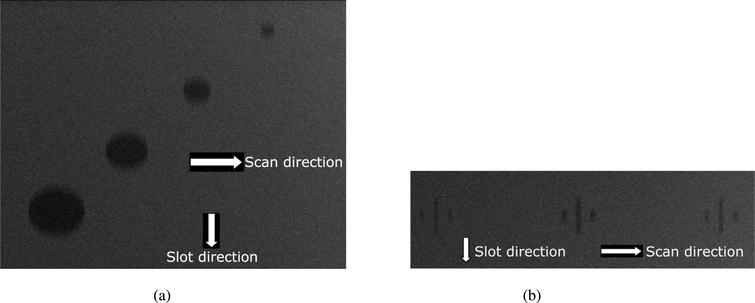
- An amorphus selenium (a-Se) flat-panel detector was modified to implement the ALER technique for slot-scan imaging. A stepping-motor driven fore-collimator was mounted in front of an X-ray tube to generate a scanning X-ray fan beam. The scanning speed and magnification were adjusted to synchronize the fan beam motion with the image line readout.
- Slot Scan function is easy-to-use and frequently used for routine radiography. And end points Check images as Fig. Slot Scan Ease-of-Use Fig. 3 shows flowcharts of the work flow for cassette radiography and Slot Scan radiography. The conventional CR long-length imaging operation is complex. It requires switching the IP and reading.
- Early studies and clinical trials show that DBT is an improvement over full field digital mammography (FFDM) because it provides the radiologist with better image quality and more information.OBJECTIVE: This paper presents a simulation system to model the performance of a slot-scanning FFDM and DBT system.METHODS: A tissue-equivalent three.
Slot scanning mammography was developed a decade ago 7 and provides one way to reduce scattered radiation in measurements. Prior implementations were designed with a moving x-ray source, stationary slit, and moving detector. The slot-scanning system evaluated in this paper was obtained through a grant from the Canadian Foundation for Innovation. The evaluation was supported through research funds by Biospace Med, the company producing the new slot-scanning device that is the subject of this manuscript.
Abstract
A desirable mammography imaging system should offer high spatial resolution, high sensitivity, a wide dynamic range and a means of limiting detected x-ray scatter. Significant scatter reduction can be obtained with a slot scan acquisition format with minimal attenuation of the primary beam. A potential receptor for digital mammography is comprised of a x-ray screen optically coupled directly to a CCD detector. The dynamic range of our current front-illuminated CCD is about 400 using factory supplied readout circuitry. The high spatial resolution of the CCD (22.4 1p/mm at Nyquist frequency) and the x-ray screen are reduced by poor optical contact at this time. X-rays which interact with the2CCD generate a moderate signal (25-30% of the optical signal) when a thin (22mg/cm2) Gd2O2S:Tb screen is used. Image segments of a Kodak breast phantom were acquired with both the screen-CCD detector and the CCD detector alone. Substantial improvements in detector performance could be achieved by utilizing a back-illuminated CCD with a slow scan, correlated double sampled readout and a structured x-ray screen. The concept of direct optical coupling with structured screens can be implemented in high energy digital scanning systems.


Slot Scanning Mammography Center
- Publication:
- Pub Date:
- January 1987
- DOI:
- 10.1117/12.966986
- Bibcode:
- 1987SPIE..767..102N